
Looking for the right direction
Definition
The Compass function is an additional function that helps locate the areas from which the alteration of muscular work most likely originates on the dental arch. By clicking on the appropriate button it will show a graph consisting of black lines superimposed on the image of a dental arch.
The upper lines are directed at the molars and those below the premolars. The more a line is covered by the graph (green, yellow, or red) the more the "work" of that muscle in that area of the mouth is altered in its performance.
There are two factors to consider for the correct interpretation of the chart:
- Shape
- Color
Form
More the vertices will be positioned near the ends of the black lines the more we will be far from a physiological situation (both in excess and in defect), indicating that there is an imbalance in muscle recruitment.
Also, the shape is important: a very narrow and elongated shape towards a specific area of the mouth will indicate that a muscle is definitely more altered than others (as in figure 2, 4 and 6), a form wider and more symmetrical it will indicate a generalized imbalance, and therefore with less specific indications of work (as in figures 1, 3 and 5).
Color
The area and the shape of the quadrilateral give information on the location of the problem, but not on the severity of the imbalance. The latter is highlighted by a color code, depending on how much the muscular work performed is far from the physiological "normal" conditions.
In particular, the color of the directional graph depends on the value farthest from normality (both in excess and in defect) that is recorded among those of all the muscles analyzed.
Three situations can be identified:
- green: situation very close to normal. It is obtained for muscular involvement values within the physiological "normal" range and suggests localized occlusal changes
- yellow: limit situation. It is obtained by values of muscular involvement that leave the previously described conditions but a probable exclusively occlusal origin is maintained. A modification of the three-dimensional occlusal structure with alteration of the vertical dimension is probably necessary.
- red: situation very far from normal. It is obtained for muscular involvement values significantly higher or lower than those foreseen by the physiological "normal" conditions. Most probably a "technical" cause is added, which did not allow a correct examination to be carried out (for example, pain in the window or door, a condition of non-oral-periodontal health or trivially a defect in the electrode). In this case it is advisable to check the raw signal and repeat the test again from the beginning.
A situation similar to Figure 1 is therefore desirable, as opposed to Figure 6, although the latter has a much smaller area and shape.
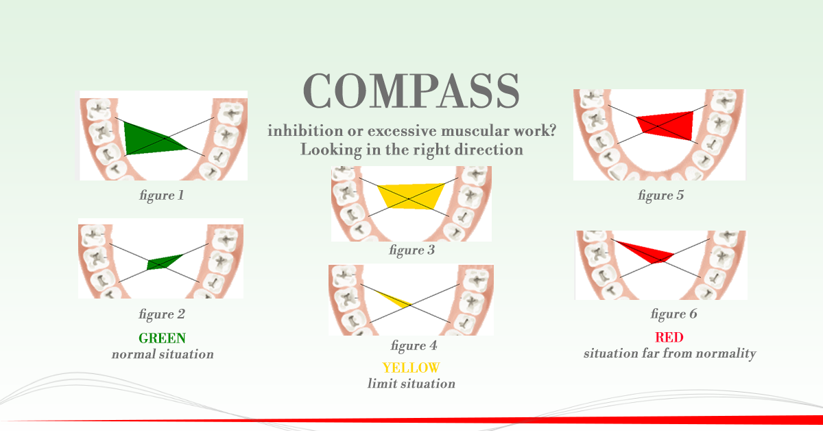
Figure 1
The temporal muscles and the right masseter have slightly altered performances (green graph therefore relatively modified values); the left masseter, on the other hand, has optimal performance values (the black line leading to the left molar areas is not covered by the green graph). The compass suggests changing the occlusal dimensions of the entire arch except the left molar area.
Figure 2
Example opposite to that visible in Figure 1; always in contexts very close to normality, the only line covered by the graph (witnessing alteration of the muscle that exerts forces in that area) is that which directs itself in the left molar area. In this condition the compass function initially suggests trying to modify the occlusal support in the left molar area. All the rest of the arch seems to be a correct three-dimensional dimension.
Figure 3
In this example all the muscles are altered in their performance significantly. It is advisable to alter the vertical dimension in the whole dental arch, also altering its three-dimensionality.
Figure 4
In this condition an important alteration of the left masseter function is shown suggesting a different support in the left molar area. Probably an alteration of three-dimensionality will be necessary.
Figure 5 and 6
They represent conditions during which serious alterations in muscular performance occurred respectively for all the muscles investigated (Figure 5) and for the right masseter (Figure 6). We recommend that you repeat the exam.